Diagnosis and Associated Risk Factors
Diagnostic assessment for retinopathy of prematurity (ROP) is by indirect ophthalmoscopy after pupil dilation using a lid speculum and with scleral depression (as-needed), facilitating an optimal peripheral retina exam.1-4 Care should be taken to use the lowest dose of dilating drops to help avoid cardiorespiratory or gastrointestinal system side effects.2,4
Risk factors for ROP:1,2,5
- Gestational age ≤30 weeks
- Low birthweight (≤1500 grams)
- Supplemental oxygen use greater than a few days or without saturation monitoring
- Poor postnatal growth
- History of hypotension requiring inotropic support, bradycardia, seizures, infection, anemia or blood transfusions
Defining Components of ROP
Zone
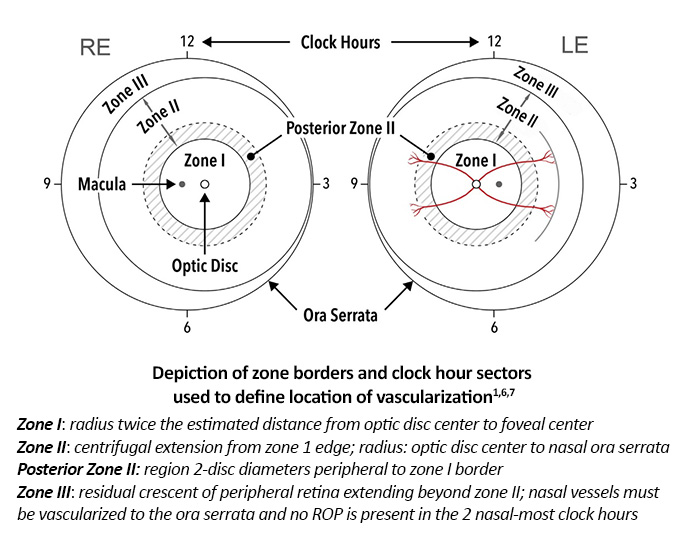
Three concentric zones centered on the optic disc were established to define location, as retinal vascular development proceeds centrifugally from the optic nerve to the ora serrata.1,6,7 Identifying the location of retinal vascularization provides not only an indication of infant maturity, but also the risk of developing ROP.6
Stage
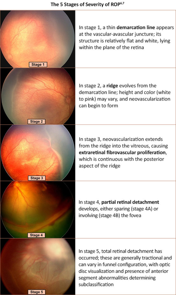
Extent of disease can be quantified by development of ROP vascular features at the junction of vascular and avascular retina.7 Since it is possible for more than one stage to be present in the eye, the most severe stage is used to classify the eye.4
Plus/Preplus Disease
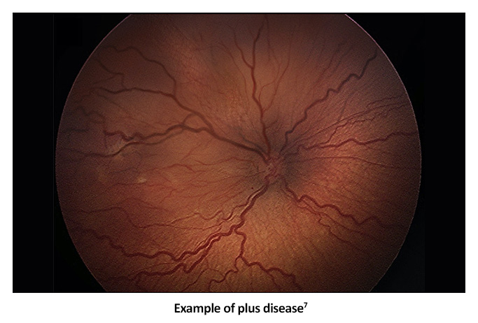
Vascular changes in zone I, specifically venous dilation and arteriolar tortuosity of the posterior retinal vessels, can be associated with severe ROP.2,6,7 Plus disease can potentially progress in severity to include iris vascular engorgement, poor pupil dilation, and vitreous haze.4,7 Plus disease is now considered to be a spectrum that begins with pre-plus disease, characterized by posterior pole vascular abnormalities that have more arterial tortuosity or more venous dilatation than considered normal.4
Severe plus disease is also a characteristic feature of aggressive ROP (A-ROP), in addition to rapid development of pathologic neovascularization without progression being observed through the typical stages of ROP.4 Also referred to as “Rush disease”, A-ROP is a rapidly progressing, severe form of ROP with vascular loops and no obvious demarcation line or ridge.4
References
- NORD Rare Disease Database. Retinopathy of Prematurity. https://rarediseases.org/rare-diseases/retinopathy-of-prematurity/
- American Society of Retina Specialists (ASRS). RetinaAtlas. Retinopathy of prematurity (ROP). https://atlas.asrs.org/article/retinopathy-of-prematurity-rop-147
- Fierson WM, American Academy of Pediatrics Section on Ophthalmology; American Academy of Ophthalmology; American Association for Pediatric Ophthalmology and Strabismus; American Association of Certified Orthoptists. Screening Examination of Premature Infants for Retinopathy of Prematurity. Pediatrics. 2018;142(6):e20183061.
- Heidar K. Retinopathy of prematurity. EyeWiki®. Last reviewed May 30, 2022. https://eyewiki.aao.org/Retinopathy_of_Prematurity
- Roybal CN. Indirect ophthalmoscopy 101. AAO Young Ophthalmologists. May 15, 2017. https://www.aao.org/young-ophthalmologists/yo-info/article/indirect-ophthalmoscopy-101
- ASRS. RetinaAtlas. Ophthalmic exam of the premature infant. https://atlas.asrs.org/article/ophthalmic-exam-of-the-premature-infant-60
- Chiang MF, Quinn GE, Fielder AR, et al. International Classification of Retinopathy of Prematurity, Third Edition. Ophthalmology. 2021;128:e51-e68.
All URLs accessed 7/1/22.
