Pathophysiology
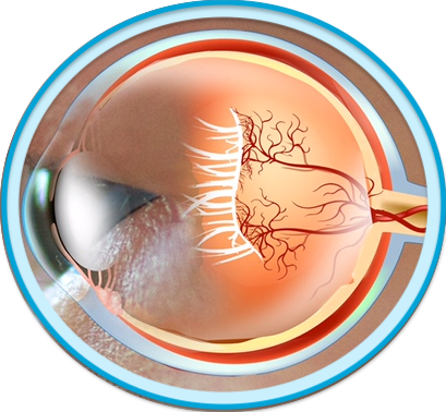
Retinopathy of prematurity (ROP) is a potentially blinding eye disorder characterized by the uncontrolled development of retinal blood vessels with disorganized branching and abnormal interconnections.1,2 ROP is most often associated with premature birth and low birth weight; the smaller a baby is at birth, the higher likelihood of developing ROP.1-3 Initially termed “retrolental fibroplasia”, ROP usually develops in both eyes and can lead to increased risk for other ocular abnormalities, including myopia, amblyopia, strabismus, retinal detachment and glaucoma.1,3
Normal retinovascular development begins approximately at 16 weeks’ gestation, with continued advancement toward the peripheral retina mostly complete at the time of full-term delivery.4 With pre-term birth, normal vessel growth may cease with incomplete vascularization of the peripheral retina.3,4
Phases of ROP pathogenesis
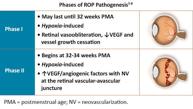
ROP occurs in two phases: hyperoxic and vasoproliferative.5 In phase I, exposure to a comparatively hyperoxic extrauterine environment leads to vasoobliteration and downregulation of vascular endothelial growth factor (VEGF) production, suppressing normal retinal vessel growth.5,6 In phase II, the postnatal maturing retina becomes hypoxic due to increasing metabolic demand, with compensatory VEGF/angiogenic factor overproduction and subsequent neovascularization between the vascular and avascular retina.5-7 Neovascularization can grow into the vitreous, resulting in hemorrhage, fibrovascular proliferation and eventual traction retinal detachment.6,7
VEGF in ROP development
Dysregulation of VEGF (specifically VEGF-A) signaling and activation of VEGF receptors (VEGFR)-1 and -2 are believed to be integral underlying mechanisms in the conversion from physiologic to pathologic angiogenesis in ROP.8 Retinal cells in animal models as well as vitreous of premature infants with advanced (vascular-active) ROP have been found to exhibit significantly higher levels of VEGF compared to controls without ROP.5
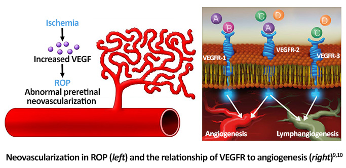
Although ROP has been previously associated with highly-concentrated supplemental oxygen use in premature newborns in the 1940s and 1950s, modern techniques facilitating precise monitoring of oxygen delivery can lower the risk for associated tissue damage.2 Alternately, post-natal light exposure has not been found to be contributory to ROP development.11
References
- Hartnett ME. Advances in understanding and management of retinopathy of prematurity. Surv Ophthalmol. 2017;62:257-276.
- NORD Rare Disease Database. Retinopathy of Prematurity. https://rarediseases.org/rare-diseases/retinopathy-of-prematurity/
- National Eye Institute. Retinopathy of prematurity. Last updated June 24, 2022. https://www.nei.nih.gov/learn-about-eye-health/eye-conditions-and-diseases/retinopathy-prematurity
- Heidar K. Retinopathy of prematurity. EyeWiki®. Last reviewed May 30, 2022. https://eyewiki.aao.org/Retinopathy_of_Prematurity
- Eldweik L, Mantagos IS. Role of VEGF inhibition in the treatment of retinopathy of prematurity. Semin Ophthalmol. 2016;31:163-168.
- Smith LEH. Pathogenesis of retinopathy of prematurity. Semin Neonatol. 2003;8:469-473.
- Neely KA, Gardner TW. Ocular neovascularization: Clarifying complex interactions. Am J Pathol. 1998;153:665-670.
- Hellström A, Smith LE, Dammann O. Retinopathy of prematurity. Lancet. 2013;382:1445-1457.
- Conrady CD, Hartnett ME. The role of anti-vascular endothelial growth factor agents in the management of retinopathy of prematurity. US Ophthalmic Rev. 2017;10:57-63.
- Sapieha P, Joyal JS, Rivera JC, et al. Retinopathy of prematurity: Understanding ischemic retinal vasculopathies at an extreme of life. J Clin Invest. 2010;120:3022-3032.
- American Association for Pediatric Ophthalmology and Strabismus. Retinopathy of Prematurity. Last updated November 4, 2021. https://aapos.org/glossary/retinopathy-of-prematurity
All URLs accessed 7/6/22.


